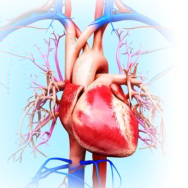
Imagine a heart the size of a walnut, its arteries and veins as slender as spaghetti. The surrounding nerves and lymphatic vessels are the width of a few strands of hair and as fragile as wet tissue paper. To repair the smallest hearts, surgeons must navigate this delicate and complex minefield.
What's more, because anatomy varies from person to person, no two surgeries are exactly the same – even among patients with the same condition. Surgeons therefore want to learn as much as possible about a patient's unique anatomy before setting foot in the operating room.
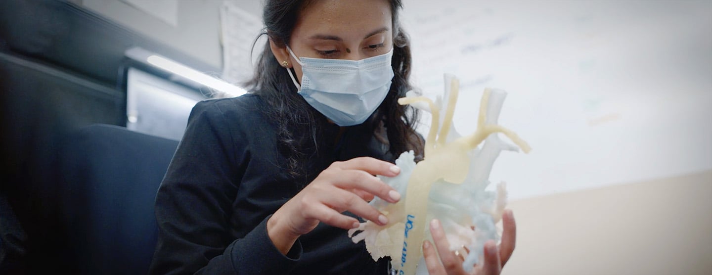
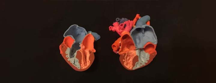
At the Pediatric Heart Center 3D+ Program, we're harnessing the power of three-dimensional technology to help surgeons do just that. With medical-grade 3D printing equipment and images taken with CT, MRI and ultrasound, we're able to produce accurate, exquisitely detailed models of patients' hearts.
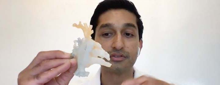
Surgeons can hold an exact replica in their hands before surgery, examining every aspect of that particular heart's structure in order to determine the best approach for repair. They can even practice procedures in advance, using models that match the texture and density of the patient's heart and that can be cut and sewn like human tissue. Or they may use virtual reality and other advanced visualization technologies to see the patient's anatomy in three dimensions while planning the surgery.
UCSF is one of only a few hospitals in the nation to offer this technology to our heart surgery team and patients, and we are the only program of our kind in the Western U.S. We also work with providers at other institutions to produce 3D models for their patients.
Benefits
Although 3D applications are relatively new in medicine, experts believe they offer advantages for patients, families and doctors.
- They may make heart procedures easier on patients because they may bring:
- A lower risk of certain errors during surgery
- Less chance of needing repeat surgeries
- Shorter procedures
- Faster recoveries
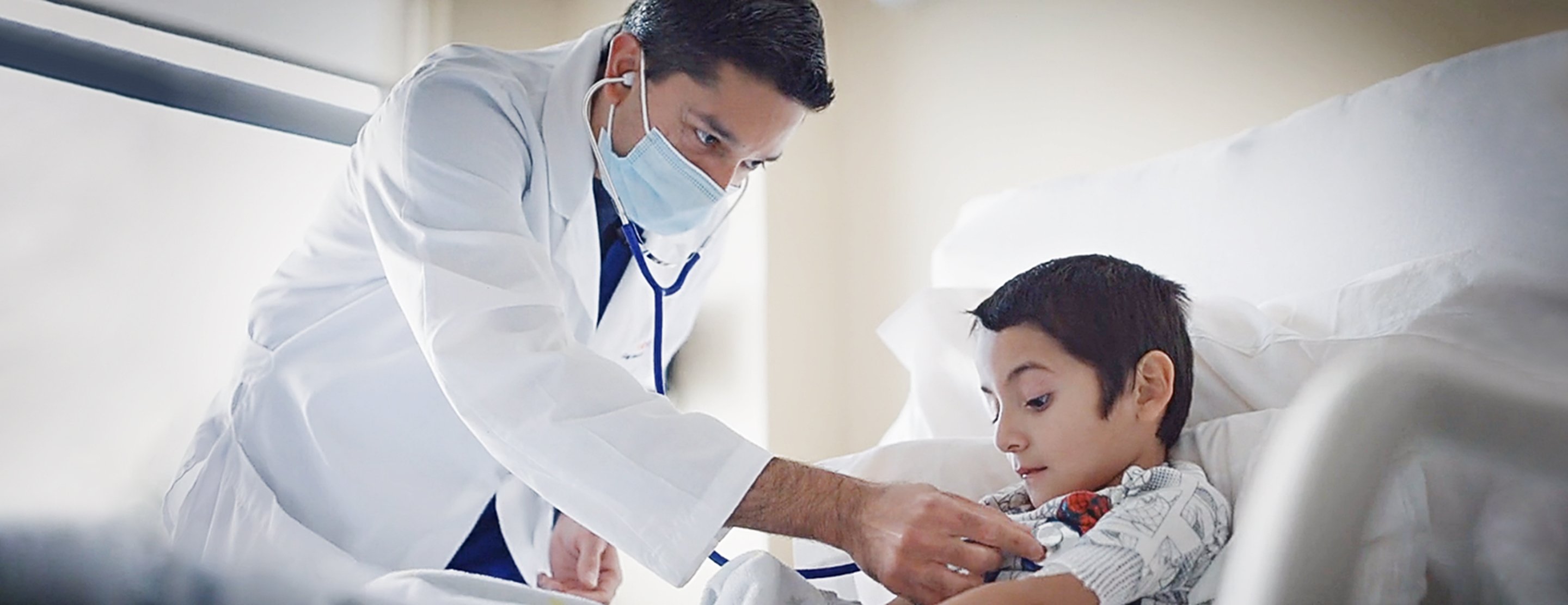
- They help families understand their children's specific heart conditions and treatment options.
- They can be used in biomedical research and to train future specialists in care for heart birth defects.
The Pediatric Heart Center 3D+ Program is part of the UCSF Center for Advanced 3D+ Technologies, which was founded in 2018 by doctors from the Pediatric Heart Center and our orthopedic and radiology departments. The team includes doctors, a dedicated biomedical engineer and image processing experts.
If you're interested in our services, please ask your child's doctor to make a referral.