


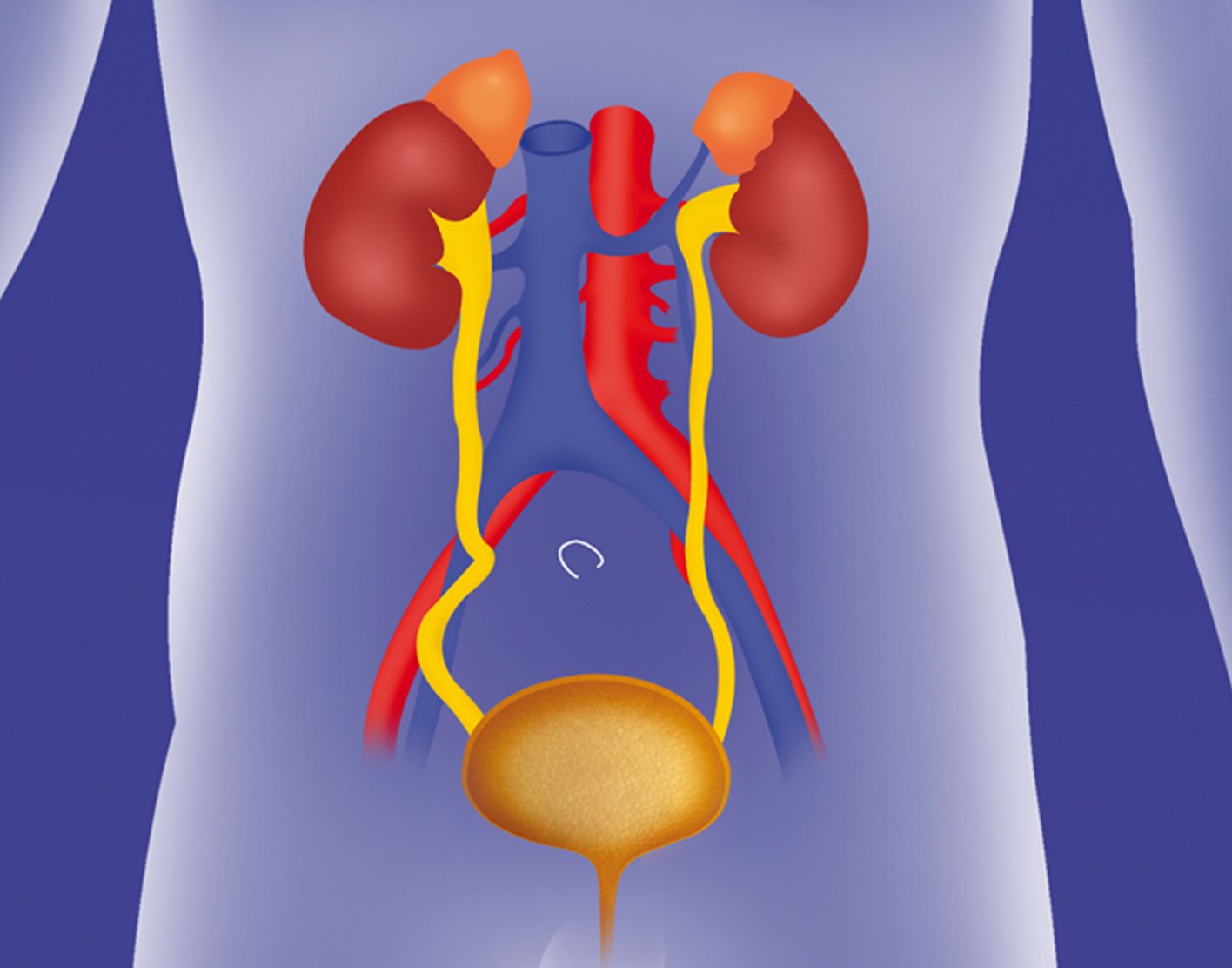
Urodynamic Testing
Urodynamics are performed in the radiology department to evaluate lower urinary system function. The purpose of this test is to evaluate the mechanism for how the urinary system works, and therefore provide accurate diagnosis and treatment of urinary system concerns. Urodynamics are a detailed version of a voiding cystourethrogram (VCUG, see Cystogram Handout) in which bladder function pressures are measured during filling, storage, and voiding (urinating). Urodynamics uses a combination of electromyogram (to evaluate for pelvic floor contractions), cystometrogram (to evaluate bladder pressures, capacity and emptying) and voiding cystourethrogram to evaluate lower urinary system function.
Urodynamics are recommended by medical providers for various reasons, including:
- To diagnose cause of urinary incontinence (accidents) if not treated by conventional measures
- If urinary symptoms do not correlate with physical exam findings
- Incomplete bladder emptying
- To determine if a medication treatment is warranted or effective,
- Spina bifida, spinal cord injuries, or other neurological conditions that may cause bladder function changes
- Prior to surgery for the urinary system, spinal, or neurosurgical interventions.
Preparation:
Urodynamics are performed in a private radiology room with an x-ray technician or nurse under nurse practitioner or physician supervision. There is no preparation for this test and no anesthesia is involved.
Please notify the technician if your child has any allergies, especially to contrast solution, or if you suspect anyone in the room is pregnant. X-rays are usually avoided during pregnancy due to the risk that radiation exposure may harm the developing fetus. If a parent would like to stay in the room to comfort their child, they will be asked to wear a lead apron to protect them from radiation exposure.
Your child may bring a special toy with them to the procedure if they would like to, and pacifiers/soothing toys are welcome. The procedure usually takes between 30 minutes to 1 hour.
Procedure:
A radiology technician or nurse will assist your child in a private radiology room. Your child will be asked to change into a gown and then lay down on a table. A large radiology machine will hang over the patient table to take radiographic images, or pictures, of your child.
The radiology nurse/technician will place three small leads, or small sticky patches attached to a cord and computer, near the rectum (anus) and legs. These are sensors to measure muscle activity.
The radiology nurse will carefully clean the urethra (the tube connecting the bladder to the outside) opening and a catheter (soft plastic tube) will be inserted into the urethra and into the bladder. The catheter is then taped in place against your child’s leg. The catheter will then be attached to tubing and filled with liquid contrast solution. A small plastic sensor will also be gently placed about an inch into the rectum and taped securely to the leg.
The radiologist or nurse/technician will arrange the machine above your child in order to take pictures of the abdomen and pelvis. The nurse practitioner or physician will start the contrast fluid infusion with the computer and the contrast will then run into the bladder and fill the bladder.
During filling of the bladder your child may be asked questions to assess if they feel the urge to urinate, when the urge is strong, and when they need to urinate. They may also be asked to cough.
Multiple radiographic images will be taken during this time.
When your child feels the urge to urinate or the bladder is full, the contrast infusion will be discontinued and your child will be able to urinate into a container. Infants will urinate spontaneously. Radiographic images will also be taken during urinating. When your child feels they have completed urinating or the infant appears to be done, a final picture is taken and the catheter is removed.
What to Expect:
After the procedure, your medical provider will analyze and discuss the results with your family.
Your child may experience some discomfort during insertion of the catheter or with beginning urination. After the procedure, there may be discomfort with the next several urination episodes and small blood spotting may be noted. If your child has continued pain and discomfort, or develops a fever or other concerns, please contact your medical provider.
Risks
- Please notify your technician and provider if you child has an allergy to iodine, as allergic reaction may occur.
- There is a small risk of urinary tract infection after procedure. Please notify your provider for any signs/symptoms of infection, including pain with urination, urinary frequency, discomfort, or fever.
- There is ionizing radiation exposure with this test. Therefore a pediatric facility familiar with latest recommended technique and minimal radiation exposure is recommended for this procedure.