


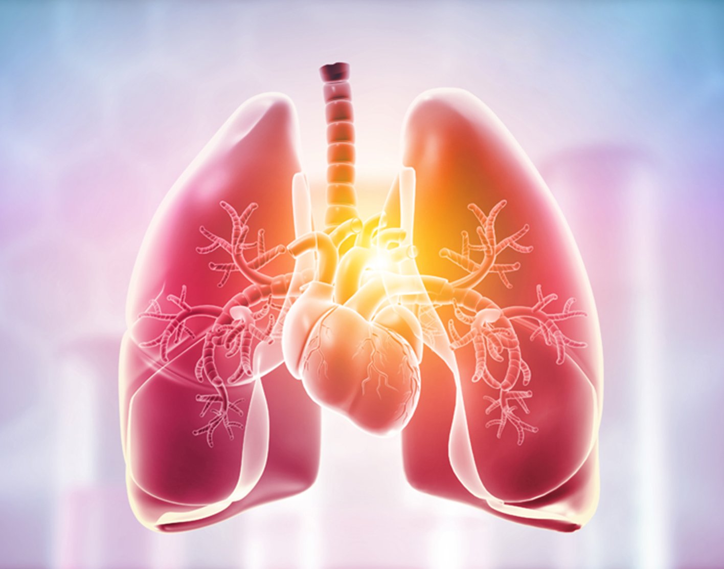
Lung PET scan
Definition
A lung positron emission tomography (
Unlike magnetic resonance imaging (
Alternative Names
Chest PET scan; Lung positron emission tomography; PET - chest; PET - lung; PET - tumor imaging; PET/CT - lung; Solitary pulmonary nodule - PET
How the Test is Performed
A PET scan requires a small amount of tracer. The tracer is given through a vein (IV), usually on the inside of your elbow. It travels through your blood and collects in organs and tissues. The tracer helps the doctor (radiologist) see certain areas or diseases more clearly.
You will need to wait nearby as the tracer is absorbed by your body. This usually takes about 1 hour.
Then, you will lie on a narrow table, which slides into a large tunnel-shaped scanner. The PET scanner detects signals from the tracer. A computer changes the results into 3-D pictures. The images are displayed on a monitor for your doctor to read.
You must lie still during test. Too much movement can blur images and cause errors.
The test takes about 90 minutes.
PET scans are performed along with a CT scan. This is because the combined information from each scan provides a more complete understanding of the health problem. This combination scan is called a PET/CT.
How to Prepare for the Test
You may be asked not to eat anything for 4 to 6 hours before the scan. You will be able to drink water.
Tell your health care provider if:
- You are afraid of tight spaces (have claustrophobia). You may be given a medicine to help you relax and feel less anxious.
- You are pregnant or think you might be pregnant.
- You have any allergies to injected dye (contrast).
- You take insulin for diabetes. You will need special preparation for the test.
Tell your provider about the medicines you are taking. These include ones bought without a prescription. Some medicines can interfere with the test results.
How the Test will Feel
You may feel a sharp sting when the needle containing the tracer is placed into your vein.
A PET scan causes no pain. The table may be hard or cold, but you can request a blanket or pillow.
An intercom in the room allows you to speak to someone at any time.
There is no recovery time, unless you were given a medicine to relax.
Why the Test is Performed
This test may be done to:
- Help look for lung cancer, when other imaging tests do not give a clear picture
- See if lung cancer has spread to other areas of the lungs or body, when deciding on the best treatment
- Help determine if a growth in the lungs (seen on a CT scan) is cancerous or not
- Determine how well cancer treatment is working
Normal Results
A normal result means the scan did not show any problems in the size, shape, or function of the lungs.
What Abnormal Results Mean
Abnormal results may be due to:
- Lung cancer or cancer of another area of the body that has spread to the lungs
- Infection
- Inflammation of the lungs due to other causes
Blood sugar or insulin level may affect the test results in people with diabetes.
Risks
The amount of radiation used in a PET scan is low. However, it is more than twice as much as most CT scans. Also, the radiation does not last for very long in your body.
Women who are pregnant or are breastfeeding should let their provider know before having this test. Infants and babies developing in the womb are more sensitive to the effects of radiation because their organs are still growing.
It is possible, although very unlikely, to have an allergic reaction to the radioactive substance. Some people have pain, redness, or swelling at the injection site. This soon goes away.
References
Deroose CM, Dooms C. Positron emission tomography. In: Broaddus VC, Ernst JD, King TE, et al, eds. Murray and Nadel's Textbook of Respiratory Medicine. 7th ed. Philadelphia, PA: Elsevier; 2022:chap 25.
Padley SPG, Popat S, Devaraj A. Pulmonary neoplasms. In: Adam A, Dixon AK, Gillard JH, Schaefer-Prokop CM, eds. Grainger & Allison's Diagnostic Radiology. 7th ed. Philadelphia, PA: Elsevier; 2021:chap 8.
Review Date: 31/12/2023
The information provided herein should not be used during any medical emergency or for the diagnosis or treatment of any medical condition. A licensed physician should be consulted for diagnosis and treatment of any and all medical conditions. Call 911 for all medical emergencies. Links to other sites are provided for information only -- they do not constitute endorsements of those other sites. Copyright ©2019 A.D.A.M., Inc., as modified by University of California San Francisco. Any duplication or distribution of the information contained herein is strictly prohibited.
Information developed by A.D.A.M., Inc. regarding tests and test results may not directly correspond with information provided by UCSF Health. Please discuss with your doctor any questions or concerns you may have.