


Learning never stops
Our classroom and bedside classes help kids get credit and keep learning during treatment.
See our school program


Here to serve you
Explore our network of care for kids, from the tiniest to teens and young adults.


World-class docs
We'll help you find the best provider for your child.


Stress-free visits
Accommodations. Admission. Discharge. Procedure prep. We've got you covered.


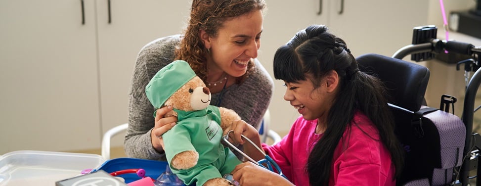
Extraordinary kid care
Our specialists handle conditions ranging from the common to the most rare.


Referrals made easy
Contacts and resources to get your patients to our pediatric specialists


Best in Northern CA
We're ranked #1 in pediatric cancer, heart surgery, cardiology & more.


Lungs are normally divided into sections called lobes; three on the right and two on the left. A congenital cystic adenomatoid malformation (CCAM) is an abnormality of one or more lobes in which the lobe forms as a fluid-filled sac, called a cyst, which does not function as normal lung tissue. CCAMs develop with equal frequency on either side of the lung, but rarely occur on both sides.
Most CCAMs either shrink or are small enough not to cause a problem. They are almost always benign, although in rare cases, they become cancerous later in life. The cyst also can become infected and cause pneumonia. For this reason, it's generally recommended to remove CCAMs after birth.
But exceptionally large CCAMs can result in serious and potentially fatal problems in fetuses.
Most cases congenital cystic adenomatoid malformation (CCAM) are diagnosed via prenatal ultrasound, before the child is born. However, the first sign of CCAM is often a pregnant mother who measures too big for her due date because there is too much amniotic fluid. This is due to the CCAM pushing on the heart and esophagus of the fetus, preventing the fetus from swallowing amniotic fluid.
A large CCAM can cause a condition called hydrops — accumulation of fluid in the skin, chest, or abdomen that reflects severe heart failure — as it presses against the heart and makes it work harder to circulate blood. About 10 percent of all fetuses with CCAM develop hydrops. Untreated, a fetus with hydrops and CCAM usually will not survive.
The mass can be so large that it limits lung development and causes pulmonary hypoplasia, or small lungs. The CCAM can also push on the heart and the esophagus of the fetus, preventing the fetus from swallowing amniotic fluid. This can result in the mother suffering from polyhydramnios, or too much amniotic fluid.
A congenital cystic adenomatoid malformation (CCAM) is usually diagnosed before birth during a pregnancy ultrasound, which will show a bright mass in the fetus' chest. The size of the mass varies with each fetus and can change dramatically throughout the pregnancy. The ultrasound may also show displacement of the heart from its normal position, a diaphragm that is flat or pushed downward, or the absence of visible lung tissue.
Fetuses who don't have hydrops — fluid accumulation that indicates heart failure — when CCAM is first detected must be followed closely with frequent ultrasounds, to check for the development of hydrops.
Most babies with CCAM are treated with surgery soon after birth or several months later, depending on the severity of the CCAM. A small group of severe cases may be treated before birth with fetal intervention.
Most infants born with a CCAM have no symptoms at birth, although occasionally an infant may have difficulty breathing and will require oxygen and the use of a machine called a ventilator.
Infants with no symptoms at birth can go home after a few days in the hospital and return at 3 months of age for a CT scan. An operation to remove the CCAM may be scheduled after the pediatric surgeon and radiologist (a physician trained to read X-rays) review the results. CCAM removal is generally recommended because of the risk of lung infections and cancerous transformation later in life.
In most infants, the operation is done with tiny telescopic instruments though several very small incisions, instead of a single large chest incision. Your baby will stay in the hospital two to three days but probably will be released as soon as he or she is breathing easily, takes formula or breast milk well and is comfortable on pain medication given orally.
Fetal intervention is offered only when there is evidence of heart failure in the fetus. The procedure involves an operation for the mother, like a Caesarean section, in which a fetal surgeon removes the CCAM from the fetus' chest.
UCSF Benioff Children's Hospitals medical specialists have reviewed this information. It is for educational purposes only and is not intended to replace the advice of your child's doctor or other health care provider. We encourage you to discuss any questions or concerns you may have with your child's provider.

Top 10 in the nation for neonatology
Ranked among the nation's best in 11 specialties
Learning never stops